


Reimagining Skin Health
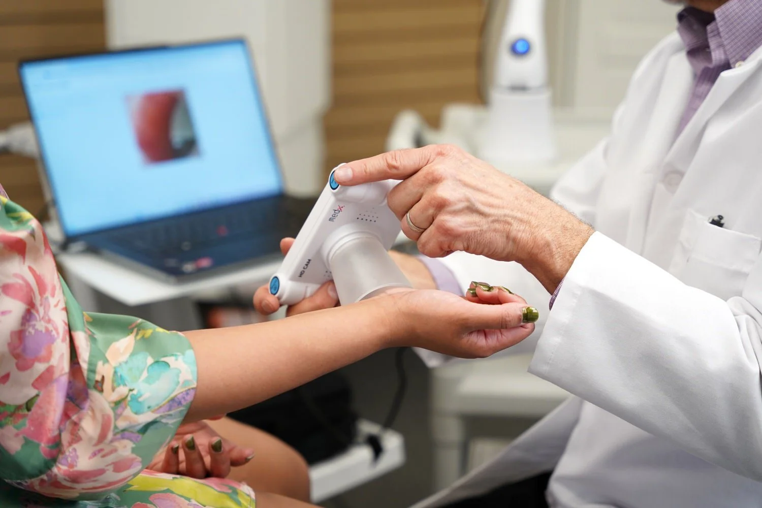
MedX uniquely delivers real-time, non-invasive measurement of skin chromophores, the biological markers in the skin that interact with light and reveal what’s happening beneath the surface to provide objective, data-driven clinical proof of sub-dermal skin biomarkers including collagen, melanin, and hemoglobin.
Born from a mission to eradicate skin cancer by making early, accessible detection the global standard of care, MedX reveals subsurface vascularity, pigmentation, and sun damage that traditional aesthetic imaging cannot detect.
This is skin health — measured, proven, and monitored over time.
Objective Skin Health – Beyond the Surface
MedX replaces subjective visual inspection with quantitative, reproducible skin biomarkers —structured for longitudinal analysis across healthcare providers, clinical research organizations, and enterprise partners.
Through advanced chromophore mapping, MedX enables partners to:
Capture real-time, non-invasive subsurface biomarker data
Quantify collagen, melanin, and hemoglobin with clinical precision
Track changes over time through repeatable, standardized imaging
Enable earlier detection, study validation, and evidence-based decision-making
MedX delivers longitudinal chromophore analysis, creating a living record of skin health — creating structured skin intelligence, not just images.
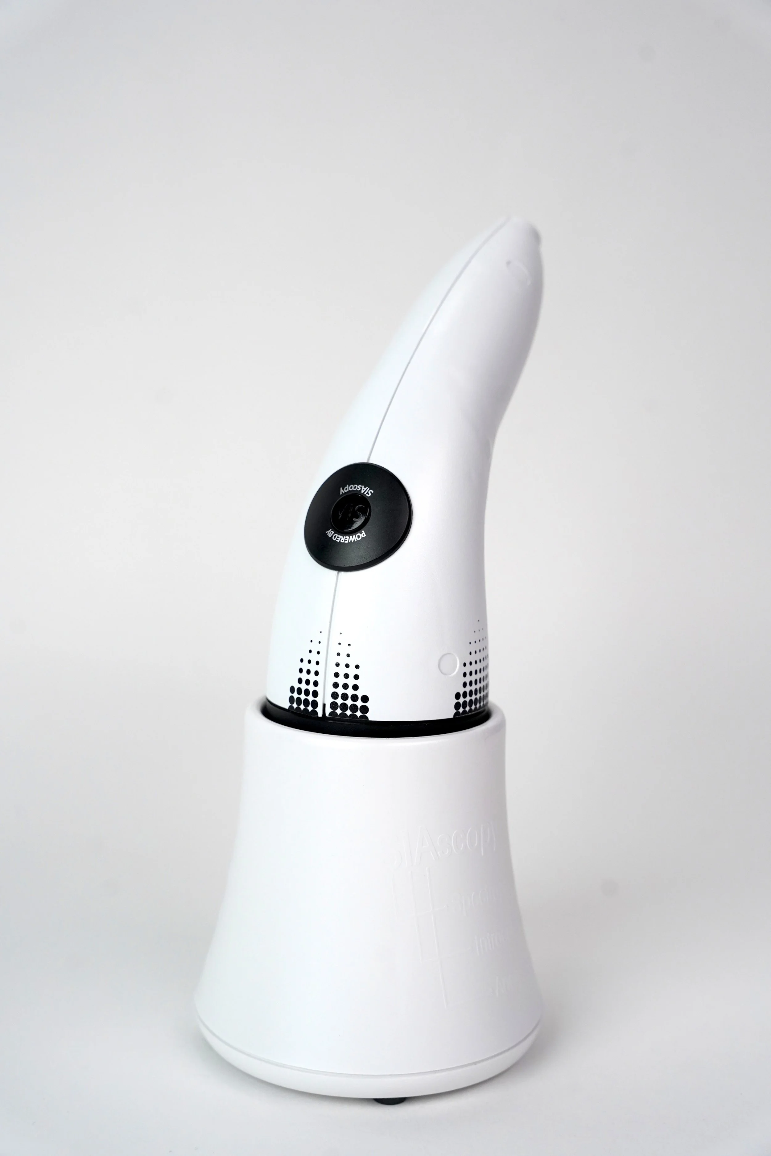
Our Products
The Choice of Professionals
-

DermSecure
DermSecure is the FDA-cleared clinical assessment and detection layer of the MedX Skin Health ecosystem, designed to make early skin cancer detection faster, more accurate, and more scalable.
-
MedX Laser Therapy
MedX’s Class IIIb photobiomodulation (PBM) laser systems deliver safe, non-invasive, drug-free therapy that stimulates cellular healing, reduces pain and inflammation, and empowers providers to improve outcomes across a wide range of clinical applications.
-
SkinSecure
SkinSecure™ is MedX’s cloud-based platform that uploads chromophore images for immediate clinical trial analysis, measuring sub-dermal (up to 2mm) changes in melanin, hemoglobin, and collagen to deliver evidence-based insight into treatment efficacy.
Built for Clinical Rigor, Global Compliance, and Interoperability
MedX was engineered from day one for regulated healthcare environments — establishing a foundation of clinical and regulatory rigor that underpins every product we build and enables us to scale with confidence.

Regulatory Approvals
FDA Cleared • Health Canada Approved • CE Accredited
Operates within ISO 13485 quality management standards and MDSAP-aligned processes.

Global Outreach
Secure, governed data infrastructure supporting deployment across 37 territories worldwide and architectured to support HIPAA and GDPR compliance.

Interoperability
Modular software architecture enabled by HL7 FHIR standards, supporting seamless integrations with:
Employer Health Rerecord Systems
Claims or Care Management Platforms
Research Databases
Enterprise Data Warehouses
CTMS or EDS Platforms
MedX delivers true functional interoperability, not siloed imaging, transforming imaging into structured, exchange-ready health data.

Proven at Scale – Trusted Worldwide
MedX is not experimental technology. It is clinically validated, operationally deployed, and globally trusted.
85,000+ images processed across the UK, Canada, Norway, the Netherlands, and beyond
14+ peer-reviewed validations supporting scientific credibility
Adopted by clinical research organizations, dermatologists, and enterprise partners
Designed to support longitudinal studies, population health initiatives, and preventive care models
This is data you can trust — at scale.
Learn more about MedX and how we’re reimagining skin health.

Who MedX Serves
MedX delivers a unified skin health intelligence layer for:
Dermatology
Healthcare Delivery Organizations
Payers and Value-Based Care Organizations
Contract Research Organizations (CRO’s)
Academic and Research Institutions
Investors
Wherever objective skin health data is required, MedX delivers.
The Future of Skin Health is Measured
MedX replaced subjective interpretation with clinical proof, longitudinal insight, and accessible detection — redefining how skin health is understood, monitored, and protected worldwide.
Learn more about MedX and how we’re reimagining skin health.